-
HOME
-
ABOUT
-
WHY CHOOSE US
-
TREATMENTS
-
FEES
-
CONTACT
-
FAQS

HEATHWOOD
DENTAL PRACTICE
- A Quick Guide To… -
TOOTH ANATOMY

Teeth can be broadly divided into the part visible above the gum, the ‘crown’, and the ‘root’ which is buried in bone, under the gum. The bone holds the tooth in place.
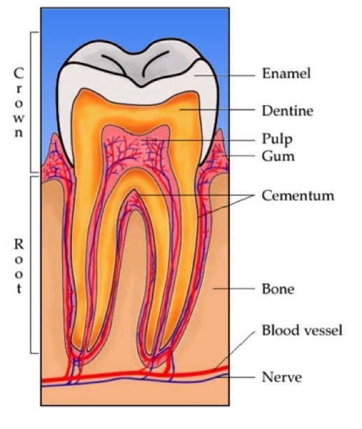
The outer layer of the crown is composed of Enamel. This whitish translucent layer is the hardest substance in the body. It is thickest on the biting surface of the tooth, and thinnest at the sides, especially near the gum edge.
Supporting the enamel, and also composing the root of the tooth is Dentine. This yellowish porous material is softer than enamel, and is perforated by many microscopic channels. Within some of these are nerve fibres, which give dentine a certain amount of sensitivity, allowing us to detect problems such as decay. When gum recession occurs, such that the dentine of the root is exposed in the mouth, these areas can become sensitive., especially but not exclusively to temperature changes (for example, hot and cold drinks).
Within the centre of each tooth, enclosed by dentine, is contained a collection of nerves and blood vessels, collectively known as the pulp. The pulp connects to the main nerves and blood vessels of the jaws through the tip of each root.
Common problems that we encounter with teeth are:


CONTACT US TO BOOK OR FOR MORE INFORMATION
This website makes use of cookies. Please see our privacy policy for details.
OK
